Complete Human Penile Scaffold for Composite Tissue Engineering: Organ Decellularization and Characterization
Complete Human Penile Scaffold for Composite Tissue Engineering: Organ Decellularization and Characterization
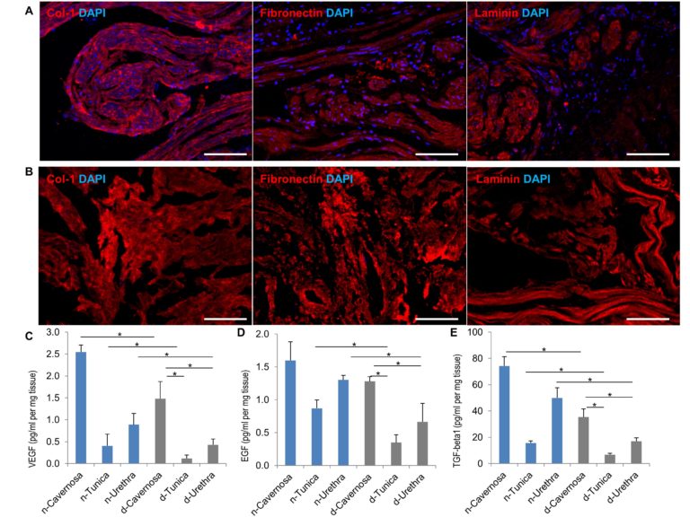
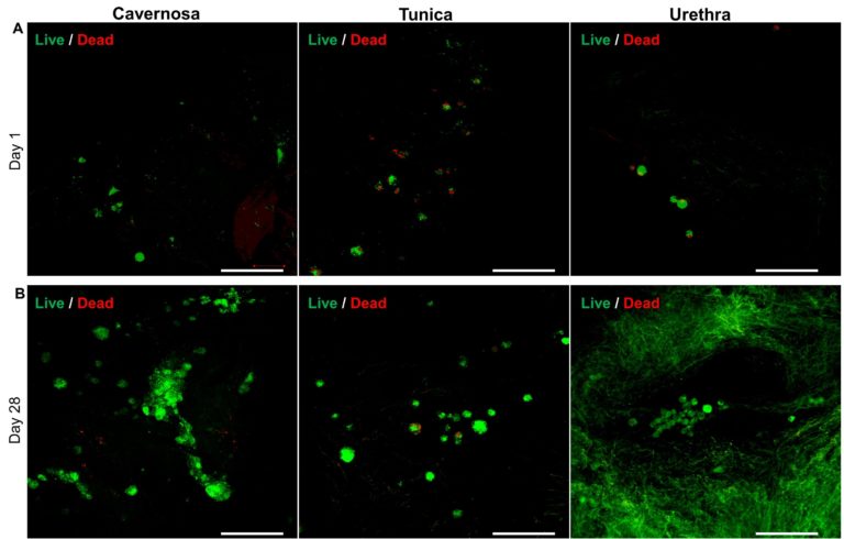
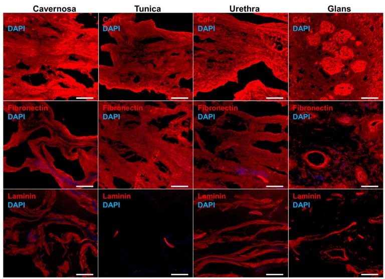
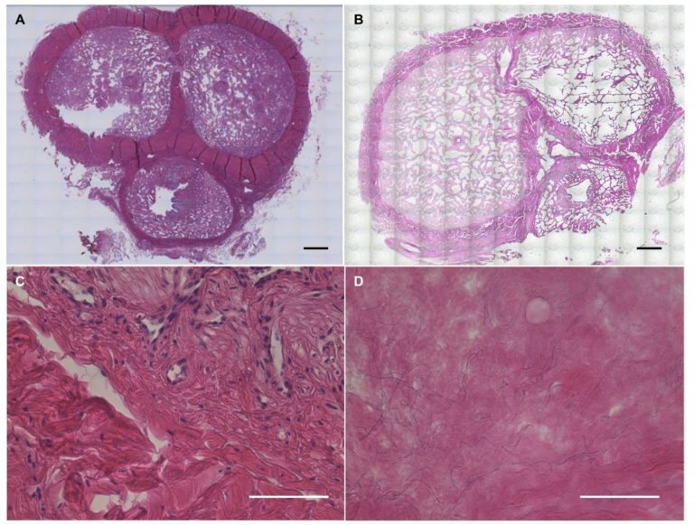
Reconstruction for total penile defects presents unique challenges due to its anatomical and functional complexity. Standard methods suffer from high complication rates and poor functional outcomes. In this work we have developed the first protocol for decellularizing whole-organ human penile specimens for total penile tissue engineering. The use of a hybrid decellularization scheme combining micro-arterial perfusion, urethral catheter perfusion and external diffusion enabled the creation of a full-size scaffold with removal of immunogenic components. Decellularization was completed as assessed by H&E and immunohistochemistry, while quantification of residual DNA showed acceptably low levels (<50 ng/mg). An intact ECM was maintained with histologic architecture preservation on H&E and SEM as well as preservation of key proteins such as collagen-1, laminin and fibronectin and retention of growth factors VEGF (45%), EGF (57%) and TGF-beta1 (42%) on ELISA. Post-decellularization patency of the cavernosal arteries for future use in reseeding was demonstrated. Scaffold biocompatibility was evaluated using human adipose-derived stromal vascular cells. Live/Dead stains showed the scaffold successfully supported cell survival and expansion. Influence on cellular behavior was seen with significantly higher expression of VWF, COL1, SM22 and Desmin as compared to cell monolayer. Preliminary evidence for regional tropism was also seen, with formation of microtubules and increased endothelial marker expression in the cavernosa. This report of successful decellularization of the complete human phallus is an initial step towards developing a tissue engineered human penile scaffold with potential for more successfully restoring cosmetic, urinary and sexual function after complete penile loss.
This paper was written by Yu Tan, Wilmina N. Landford, Matthew Garza, Allister Suarez, Zhengbing Zhou, and Devin Coon and published by Nature.


